Are they new?
All HART articles also on Substack. Please consider a PAID SUBSCRIPTION so we can continue our work. Comments are open so you can join in the conversation.
Numerous funeral directors have now reported struggling to embalm bodies because of finding white “calamari-like” clots blocking the veins. What do we know about these clots? John Campbell has interviewed a series of funeral directors who describe a decreasing incidence of such clots but claim they remain in about 20% of those who die in 2023.
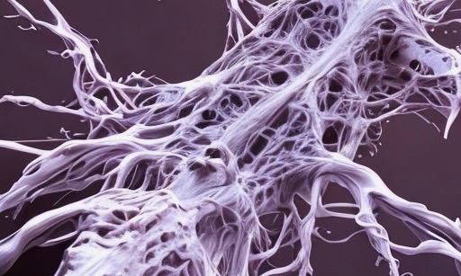
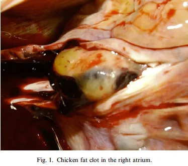
Are they just conventional post mortem clots? Although some may have formed post mortem they are not what is usually seen. Post mortem, the blood can separate into a yellow plasma fraction and a red cellular fraction and each can clot separately. The result is a soft, jelly-like, yellow clot known as a “chicken fat clot” attached to a “blackcurrant jelly” clot. The yellow colouring comes from entrapped lipids, proteins and bilirubin in the plasma. What the embalmers are describing is white throughout and has tensile strength – even before being placed in formaldehyde. Is the testimony from multiple funeral directors so easily dismissed as if they had never seen post mortem clots before?
Recently, the story has dramatically shifted into problems in the living. An experienced technician in a catheter laboratory has reported seeing extensive clots in life, which when removed appear almost like casts of the blood vessels, as shown in an interview with Dr Philip McMillan here. Although many of these clots appear redder than the post mortem clots but he claims this is because they are still coated in fresh blood.
In a follow-up interview, Dr McMillan has talked to pathologist, Professor Resia Pretorius, who again highlighted the need for proper examination of the morphology and chemistry of these clots. She had previously published on micro-clots as a result of the viral infection. Her team went on to show that, even in the absence of platelets, spike and plasma (cell free blood) make cell free clots. Spike interacts with clotting fibres – fibrinogen – to flatten them out into a pathological type of protein called amyloid which stack together to make larger structures. As Professor Pretorius says, until it is known exactly what these much larger clots are made of, it is impossible to start working out how best to treat them.
White blood clots did occur pre-covid panic – in the arterial system. The colour of these arterial clots is white because there are few red blood cells in them – it’s a sign they were made in a high flow environment. However, the funeral directors have removed these “calamari clots” from the venous system which is not a high flow environment.
There must be a different reason why these “clots” do not trap red blood cells despite being in the venous system.
Normal clots require a pro-thrombotic surface e.g. from disruption of the cells lining vein and artery walls – endothelial cells along with a chemical cascade of clotting components within the blood. There are numerous ways in which the covid vaccines can interfere with this process:
- The mRNA platform results in foreign protein expression in endothelial cells and their consequent death. This creates a pro-thrombotic surface. Furthermore, endothelial cells use heparin to keep the vessels free of clots and spike interrupts that.
- Spike results in platelet activation in mice. The receptor binding domain (included intact in the vaccine) alone “could bind platelets, cause platelet activation, and potentiate platelet aggregation.”
- It’s not just mice. Plasma from a 25 yr old woman given Moderna vaccine showed dose dependent platelet activation. The more spike protein present the more platelet activation occurred. She did not have VITT.
- The inflammatory state created post vaccine is itself pro-thrombotic – although these would likely be conventional clots.
It is hard to think of any ways in which a product COULD cause clotting that hasn’t already been shown to apply to these products.
Could it be the virus?
SARS-CoV-2 is a respiratory virus and it produces spike protein at the respiratory surface. In very sick people RNA can be detected in the blood – but not usually the whole virus. Fauci and colleagues described how it’s only viral RNA in the blood, not whole virus. What’s more, an infection lasts days to weeks only.
For the vaccines the story is VERY different. Although manufacturers claimed spike expression would last 9 days and stay in the muscle – reality says otherwise.
While there is plenty of evidence of a propensity to clot in the vaccinated, that does not explain why the clots are white. Some have suggested these “clots” are in fact endothelial cells that have sloughed off the vessel walls. However, microscopy shows they are proteinaceous and have no cell nuclei in them. Some have suggested that the protein is self-replicating like a prion disease. Others have suggested they are made of sheets of an abnormal protein called amyloid. That would certainly fit with several of their features but, oddly, no-one has reported on a simple, old fashioned microscopic technique to prove or disprove this – a congo red stain. Other investigations that are long overdue include mass spectrometry to determine the elemental makeup of the clots. Lack of funding and indeed refusal to consider such vital topics for research will continue as long as university biomedical departments receive much of their funding from the pharmaceutical industry.
Even if we knew more about their composition, questions would remain about their effect. How many of the people who die with these clots die because of them or were ill because of them?