

Usable folate can run short in the embryo while the mother’s blood folate reads entirely normal
Dr Clare Craig
This article attempts a synthesis of the science on neural tube causation.
Key points
- The story of a folate deficiency underlying all neural tube defects (NTDs) does not add up. No deficient group has ever been identified that accounts for them, cases occur right across the folate range, famine produced no rise, and the rises and declines in rates were unrelated to changes in diet.
- A mother with normal blood folate levels can still have an affected pregnancy.
- A toxin, fumonisin is the clean proof: it disables folate transport and starves the embryo while the diet and the blood are replete.
- Folic acid does have an impact on high risk women. Recurrence fell 72 percent in women who had already had an affected pregnancy on a pharmaceutical dose of 4 mg a day. The mechanism was unexplored for three decades.
- Frog experiments published this year point to folic acid working by raising the activity of an enzyme which helps convert vitamin A into retinoic acid that acts on cell proliferation in the developing brain.
- Folic acid does not prevent neural tube defects in certain circumstances e.g. in rural Bangladesh the protection from folic acid fell away as well-water arsenic levels rose and no protection remained once the levels hit 25 micrograms per litre.
A mandate to add folic acid to flour depends on three beliefs:
● that these defects arise because women do not get enough folate from their food,
● that folic acid intake will therefore prevent them and
● that it is perfectly safe for everyone else.
Myth 1: NTDs are caused by a folate deficient diet
The first point does not hold up as the only cause of NTDs. In animal models removing folate does result in NTDs but no folate deficient group has ever been identified that accounts for these defects, cases occur right across the range of folate levels including in women with the highest folate stores, famine which destroys folate intake did not cause the rate to rise, and the rises and falls in incidence are unrelated to changes in diet. Western countries that have added folic acid to food have seen neural tube defect rates plateau [HART article to come: Plateau in four countries]. I have found no series in which affected pregnancies began to fall when a folic acid policy began.
Folic acid does have an impact, particularly in high risk women. Trials with women who had already had an affected pregnancy and took 4,000 micrograms a day, a pharmaceutical dose, saw recurrence fall by 72 percent. These trials were followed by a single trial of women at ordinary risk who took 800 micrograms in a multivitamin and were compared to a control group with trace elements including manganese. The results showed a fall in affected births, but there were nine pregnancies lost for every defect prevented and its own authors noted that the whole claimed benefit could have come from the increased risk of pregnancy loss. The importance of manganese in that control arm is dealt with below.
Even if the trials in the early 1990s did prove effectiveness, the background problem was not the same. Rates were around 3 per 1,000 at the time of the trials but have since fallen and plateaued at 1.5 per 1,000. Once other causes are removed, what remains will be from causes that are harder to shift e.g. genetic susceptibility. Given the range of causative factors, it is likely that the fall was due to a reduction in some causes and if folic acid rescued those cases that does not mean it can rescue cases now that they remain at the low plateau rate.
While the pregnancy losses can explain the effect at low doses they do not explain the well demonstrated effect for the high risk women. Nobody has ever found evidence that these women are folate deficient. How did folic acid achieve that effect?
The body is complex and I do not pretend to hold the full answer. What follows sets out where the evidence stands currently. It is not a claim to be able to explain every aspect of these complex interactions. However, the evidence base has moved on significantly in thirty years and shows plainly that the deficiency story is wrong and what a better account might be.
The folate shortage hypothesis
Most doctors will tell you that folate shortage is responsible for neural tube defect formation. Replacing folates with folic acid fixes it. That is the extent of the consensus on causation.
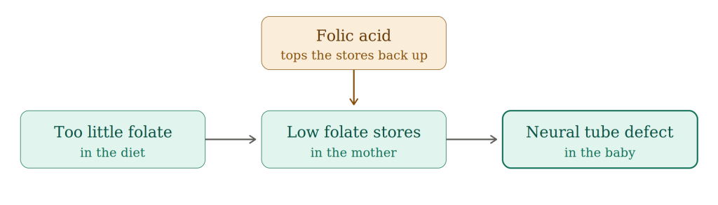
Figure 1: Model of consensus position on neural tube defect causation
The situation is more complex but this is a good place to start. Folate metabolism plays a key role in neural tube formation and understanding this helps with understanding how other toxins and drugs interact to increase the risk. The clearest demonstration of the difference between folate levels in the mother and in the developing brain is the toxin fumonisin, which blocks folate entry into the embryo causing NTDs even while the mother’s diet and blood folate stores stay replete. Before we come to that, let us step through the journey folate takes from the mother to the cells of the developing neural tube.
Mother’s blood folate stores are correlated with risk, but there is still risk in those with the highest stores, which suggests a third factor is at work. A lack of evidence of deficiency in the mother is only part of the story. Animal work points to a deficit in the embryo itself as the thing that matters, since reducing a mouse embryo’s folate supply produces these defects while the mother’s diet and blood stay normal.
Getting folate into an embryonic brain cell is the first hurdle. Folate is a charged molecule and does not simply drift across a membrane, so it has to be transported in. Most cells use the reduced folate carrier, the protein the human gene SLC19A1 codes for. The early embryo relies heavily on a second and higher affinity route, folate receptor alpha, which is found in only a few tissues, among them the neural tube lining, the placenta and the kidney. Mice lacking either entry route for folate die in the womb. If they are given sufficient folate to survive they are then born with neural tube defects. The placenta is poor at folate transport but in any case it is not in place during the critical first four weeks when the neural tube forms. Until then folate arrives by a chain of handovers from the mother to the uterine gland to the yolk sac to the embryo.
Folic acid needs to be processed before it can be used. The folates in food are already in a reduced form and can be used, while folic acid is oxidised and must first be reduced by dihydrofolate reductase before it can be used. Human liver is painfully slow at that conversion, working at under 2 percent of the rate a rat manages. Rodents were the model for the animal safety testing.
Once inside the cell and converted, folate supports three pathways that the developing neural tube, which has to roll up and fuse together, depends on. Gene switching: folates methylate genes, which allows the edges of the neural plate to align. Cell replication: folates supply the purines for cell division, so the plates grow large enough to meet each other. Growth limitation: retinoic acid holds the growth of the plate in check so that it stays the right size for fusion. Too little of it and the cells multiply too fast, the plate over-expands and its edges finish too far apart to meet. Closure of the neural tube needs retinoic acid to be within a tight range, and too much of it causes these defects as well as too little. An excess of retinoic acid is the standard laboratory method of producing these defects, in one experiment in 79 percent of mouse fetuses.
What matters in the first 28 days is therefore not how much folate the mother has but how much gets into the developing brain cells and the interactions when it arrives. Folates impact on three different pathways:
● Gene switching, which requires methylation
● Cell replication, which requires folate derived purines
● Growth limitation, which requires retinoic acid from vitamin A
Gene switching begins with the MTHFR enzyme making 5-methyl-THF, which hands its methyl group to homocysteine to make methionine, and methionine is then converted into S-adenosylmethionine, the methyl donor for DNA and histones that allows genes to be switched on and off. It is this pathway that the heavy metals interfere with. Cell replication uses folates in the form of 10-formyl-THF to make the purines needed for DNA and RNA. Growth limitation runs through ALDH1L1, an enzyme of folate metabolism that competes for that same 10-formyl-THF. It has a second and quite separate job of turning retinaldehyde from vitamin A into retinoic acid which is critical here because it is needed for successful neural tube formation. Where there is a propensity to have a neural tube defect, folic acid helps by increasing the amount of the enzyme in the key area which makes the retinoic acid.
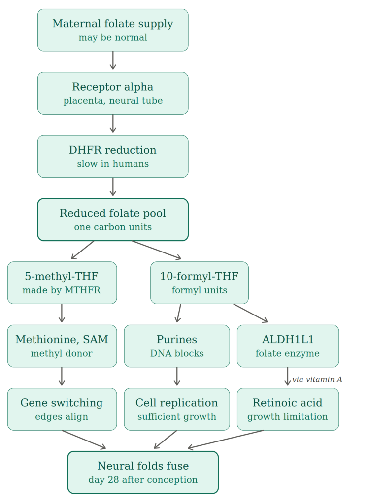
Figure 2. The pathway with nothing interfering with it.
A failure of any link in that chain can result in a neural tube defect even when the mother’s blood folate level stays normal.
Although it is slow, once folic acid has been processed it does increase folate stores, since folate is what is left after folic acid is reduced. For thirty years the assumption has been that folic acid, by increasing folate stores, could replace lacking folate and let the three pathways above flow freely. In July 2026 a quite different mechanism was demonstrated, at least in the model used. In frog embryos, folic acid could only prevent neural tube defects when the ALDH1L1 enzyme was present and there was sufficient vitamin A to make the retinoic acid, and deleting the gene abolished the protection entirely. The human version of the enzyme made retinoic acid in the test tube. This is an important demonstrated mechanism [HART article to come: How folic acid works]. Whether or not it alone can explain the result of the human trials is yet to be determined.
Myth 2: folic acid works by replacing missing folate
Folic acid is a drug but it is eventually metabolised into folate the body can use and does increase folate stores. Even women with the highest exposure to folic acid still are at risk of NTDs. That is because the causes are more complex than simply folate deficiency.
There is more than one factor that can interrupt neural tube development. Folic acid can overcome the harm in many of these, but just because it treats the problem does not mean that a lack of folate was the problem it was treating. These causes are supported by very different quality evidence. Valproate has the strongest evidence: taken in early pregnancy it produces spina bifida in one to two percent of exposed babies, a ten to twenty fold increase. Diabetes present before pregnancy carries a pooled odds ratio of 2.24 and a raised maternal temperature in early pregnancy 1.92. Pesticide exposure is suggested by systematic review but the evidence is weaker. Arsenic is a case of its own and is dealt with below, because its effect shows up only in combination with folate.
Toxins
An epidemic of neural tube defects [HART article to come: Case studies in NTD causation] along the Texas border with Mexico was traced to a toxin called fumonisin, produced by a mould that grows on maize. Fumonisin blocks the enzyme that builds sphingolipids, the fats that keep folate receptor alpha intact on the cell membrane. Cutting those fats by around 40 percent almost completely blocked folate transport through that receptor. The toxin therefore starves the embryo of folate while the diet and the blood stores stay replete. Folic acid reduced the affected proportion of embryos in fumonisin poisoned mice from 79 percent down to 50 percent. Replacing the sphingolipid, which corrects the obstruction rather than pushing past it, reduced it to 5 percent.
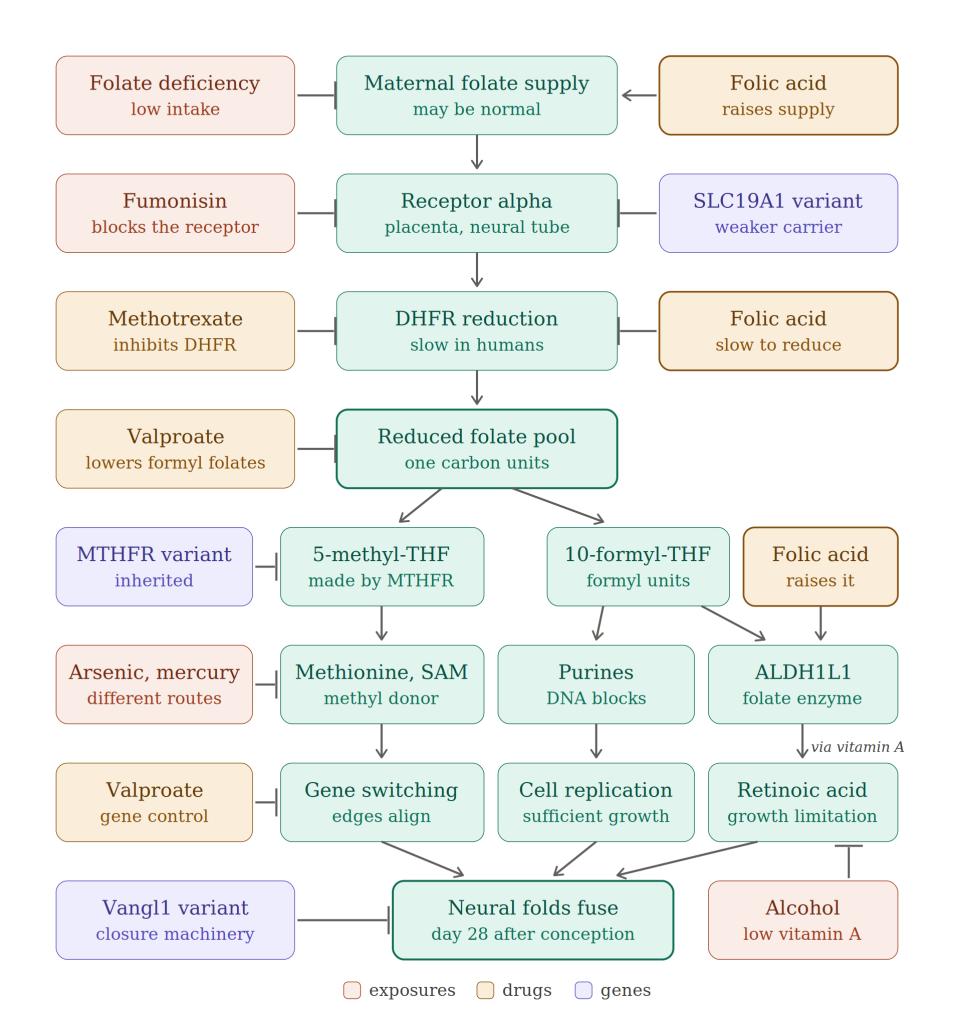
Figure 3. The folate neural tube formation pathway, with every known obstruction marked.
Autoimmune
As well as fumonisin damaging folate receptor alpha, the receptor can be blocked directly by autoantibodies raised to it. Children with cerebral folate deficiency have such antibodies, and the result is low folate levels in the cerebrospinal fluid despite entirely normal folate in the blood. However, they do not have neural tube defects, because the antibodies appear long after the tube has closed. In rats, injecting antibodies to the folate receptor when the neural folds are forming caused death of every embryo at the higher doses and about half at slightly lower ones. The survivors had growth and central nervous system defects. Folinic acid which enters cells even with a blocked folate receptor alpha, prevented the damage from the smaller doses. What the syndrome proves is that a blockage at the folate receptor alpha can result in the brain being starved of folate while the blood levels are perfectly normal.
Heavy metals
The body disposes of arsenic by adding methyl groups which are supplied from the same folate metabolism pathway that supplies methyl groups for gene switching. Its presence therefore increases the pressure on the folate pathway. In a randomised placebo controlled trial in folate deficient Bangladeshi adults, 400 micrograms of folic acid a day for twelve weeks increased the proportion of arsenic that was fully methylated and lowered total arsenic in the blood. Folate status accounts for how well arsenic is cleared but clearing arsenic puts pressure on the folate pathway.
If competition for methyl donors matters, arsenic should do more harm where the methyl supply is already marginal, and that is the pattern. In rural Bangladesh, where almost no mother takes periconceptional folic acid, more arsenic in the well water meant less benefit from folic acid, and above 25 micrograms per litre it gave no protection at all. In northern China, placental arsenic above the median carried a risk of 1.68 among the women taking no folic acid and no measurable risk among those who did. Bangladeshi women in the UK have an almost threefold increased risk [HART article to come: NTDs in migrants], which could come from exposure as children, given that 74 percent of UK mothers of Bangladeshi ethnicity in one study were born there, or from continuing exposure through imported rice.
Arsenic is an established experimental cause of these defects in animals. When given orally to mice arsenic produced neural tube defects with a clean dose response. Older work in mice and hamsters found the same defect. Some rat and rabbit studies found nothing but these are outnumbered by animals where developmental harm was demonstrated.
Folate is key because mice lacking one of the folate binding proteins were more susceptible to arsenate than wild type mice and decreasing folate intake raised the risk further. Arsenic metabolism was not changed in these mice only folate.
There is strong experimental evidence that another heavy metal, mercury, causes neural tube defects. Exposing pregnant mice orally during the critical days of closure raised the rate of neural tube defects with no extra maternal deaths or embryo loss, and the mechanism was a failure of gene switching. Mercury was blocking the key step in the methylation pathway with methionine building up in the exposed embryos while its product S-adenosylmethionine fell. Arsenic consumes the methyl donor and mercury obstructs its manufacture. In both cases the methylation pathway suffers. In humans, placental mercury levels were related to risk, with women who had levels above the median having more than an eight fold risk of a neural tube defect. There was also a dose response relationship.
In Britain, 20th century mercury exposure came from both medicines, up until 1950s, and fillings. Exposure of mothers to the former could still affect births into the 1970s and 1980s. Dental amalgam was about half metallic mercury. Cord blood mercury correlates with the number of fillings a mother has and is related to how recently they were filled. At autopsy mercury has been found in fetal liver and kidney and in infant cerebral cortex in proportion to the number of the mother’s fillings. Hot drinks may increase risk of mercury release from fillings [HART article to come: What caused the NTD epidemics in Ireland and Britain]. The incidence of neural tube defects in Britain fell over the same decades as mercury exposure in the mothers did. The majority of the drop in neural tube defects between 1964 and 2004, was due to a reduction in affected pregnancies with an increase in terminations accounting for the remainder.
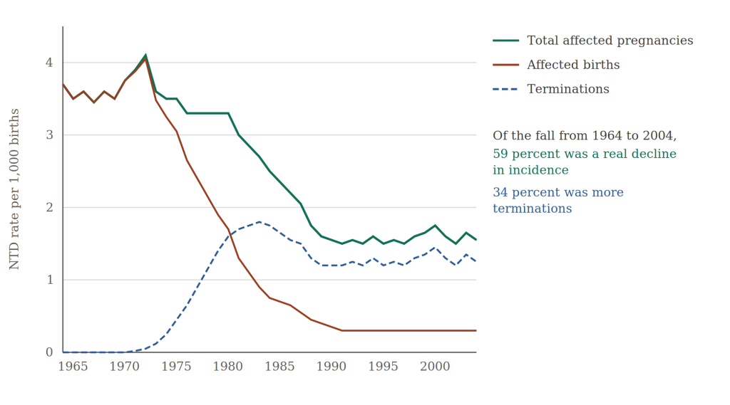
Figure 4: Decline in neural tube defect rates in Britain
There is counter-evidence but it is flawed. A Norwegian cohort of 69,474 pregnancies found no association between fillings and malformations generally but did not look at neural tube defects specifically. A study of dental nurses who mixed amalgam by hand showed no excess of spina bifida but it was too small to show any rise less than a doubling in risk.
Finally manganese, another heavy metal, has an association with these defects supported by the same 2023 systematic review that supported mercury. Manganese was included at low dose in the control arm of the only randomised trial of periconceptional folic acid in women at ordinary risk. If manganese introduces its own risk then the control was not inert and the apparent benefit may have been inflated.
Drugs
The chemotherapy and anti-inflammatory drug methotrexate inhibits DHFR (dihydrofolate reductase) directly. This is the enzyme that converts folic acid into a natural form and it is already very slow in humans. Methotrexate is known to cause birth defects. Trimethoprim, the common urinary antibiotic also blocks this enzyme and results in a fivefold increased risk of birth defects. The anti-epileptic drug, valproate, inhibits folates entering the methylation pathway and separately blocks an enzyme, histone deacetylase, needed for gene control.
Genetic variants
At least three genetic variants play a role in neural tube defect risk. The MTHFR C677T variant is found in about 45 percent of Europeans and produces an enzyme that has roughly two thirds of normal activity in those with one copy and only about a third in those with two, which is around 10 percent of Europeans. A mother with at least one copy was 1.30 times as likely to have an affected pregnancy as a mother with none and a mother with two copies 2.02 times as likely.
Mothers with a variant in SLC19A1, the gene that codes for a reduced folate transmembrane transporter had around a 55 percent higher risk of an affected pregnancy than mothers without this variant. That particular transporter is expressed in the developing neural tube and mice lacking it either die in the womb, or are born with neural tube defects if given enough folate to survive.
Finally there are the genes which code for the machinery that closes the neural tube, the planar cell polarity genes. In mice, disrupting any of more than 200 genes produces neural tube defects. In humans no single causative gene has been found. There are rare variants in certain genes that are more common in patients with spina bifida and anencephaly, but they account for a small minority of overall cases. Either way any NTDs caused by genes which are required for the physical act of closing the neural tube will not be prevented by changes to diet.
Figure 3 shows that a shortage of folate in the diet is only one of many ways in which the complex processes needed for neural tube closure can be disrupted. Some of those obstructions act on transport, some on gene switching, some on growth limitation and some on the closing machinery itself.
Myth 3: Folic acid is perfectly safe
Folic acid is by any definition a drug and all drugs have side effects. The NHS website lists those who should be careful of exposure including anyone with cancer, a heart stent or B12 deficiency. The latter is common and when undiagnosed the anaemia that ensures a patient sees a doctor and gets treated is masked such that permanent nervous system damage carries on unnoticed. The NHS also lists a long series of potential drug interactions. With these, dosages could be adjusted but the dosage of folic acid taken in each day is likely to vary widely depending on the menu. Once folic acid is in flour avoiding it becomes very hard.
There is also a risk of new cancers with a clear cancer signal for exposures below 1mg a day i.e. fortification level exposure. Higher exposures do not have a cancer risk. As with all drugs, the effects differ by dose.
The adverse effects exist because the enzyme that converts the drug into something usable is very slow such that unmetabolized drug builds up in the blood. In the UK 94 percent of elderly people who had fasted for 24 hours had unmetabolised folic acid in their blood. In the USA, with fortification, 95 percent of a random sample had detectable unmetabolized folic acid in their blood. Rodent studies need to use very high doses to produce this same effect of having unmetabolised folic acid in the blood. These studies demonstrate changes to immune function, susceptibility to infection and cancer [HART article to come: Three things high folic acid does to animals].
Children are at greatest risk of having a disproportionate intake and the government’s own modelling assumes 4.4 percent of one to three year olds will exceed the estimated age specific upper limit.
There is also clear evidence that there are negative effects even for pregnant women with higher rates of pregnancy losses, multiple births and gestational diabetes and consequences for their unborn babies by way of increased risk of atopic dermatitis, otitis media, asthma and wheezy bronchitis.
Conclusion
Neural tube closure depends on a complex network of processes: folate transport into the embryonic brain, gene switching, cell replication, growth limitation and the physical closure machinery. Different insults break different parts of it. Fumonisin is the pure case of embryonic brain cells starved of folate while the mother has plenty. There are plenty of other exposures where plentiful maternal folate has no impact on outcome. Mercury obstructs the making of the methyl donor, arsenic consumes it and some genetic variants act on the final stage of the pathway. The evidence therefore does not support a simple model in which neural tube defects are mainly the consequence of too little folate in the mother’s diet, with folic acid replacing what is missing. Folic acid can sometimes push a broken part of the network back towards working, and in a woman at high risk that is an intervention worth considering, but it acts as a drug rather than as a nutrient replacement.
The case for medicating an entire population has been built on the premise that folic acid is a simple nutrient that can help these babies and merely safely feed the remaining population. The decision to recommend all pregnant women take folic acid failed to consider the complex causes of neural tube defects and the decision to medicate the whole population has failed to consider the safety concerns.